BASE OF THUMB ARTHRITIS
Arthritis Conditions Osteoarthritis Base of Thumb Arthritis
Wrist Arthritis PIP Arthritis DIP Arthritis Base of Thumb Arthritis
Base of Thumb Arthritis
Management of Thumb Arthritis
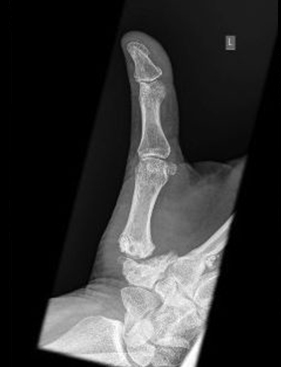
The base of thumb joint is usually the first joint that becomes arthritic and is the most common form of arthritis in the body. This refers to the specific joint between the trapezium bone and the first metacarpal of the thumb and is located at the base of the thumb. Usually, this arthritis is idiopathic and there is no real clear understanding of its pathology. There is a predilection more commonly in females at a younger age and this might be secondary to ligament laxity that results in a typical joint incongruency.
This is the most disabling form of arthritis of the hand, as 60 % of the hand function involves pinching between the thumb and the index finger. Any force that goes down the tip of the thumb is multiplied about 10 times through the first carpo-metacarpal joint.
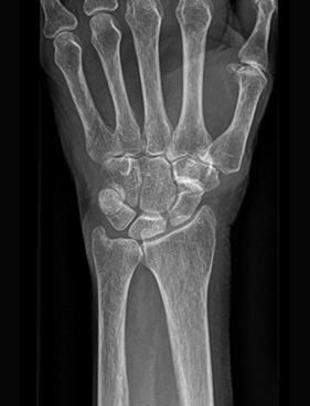
The principle of osteoarthritis is that it is a loss of cartilage between the joint surfaces. This is diagnosed clinically when the joint is examined and there is a sense of crepitus or grinding in the joint, meaning it is not a smooth surface due to the loss of the cartilage. Radiologically, there is no space between the bones. Cartilage is not visible on x-rays, and therefore, if there is bone on bone contact on the x-ray, it means there is no more cartilage left. In a chronic situation, there is loss of joint surface with secondary osteophytes and deformation of the joint. There is also a sclerotic line (abnormal hardening or thickening of bone), on the bone ends, representing a change of the bone.
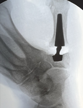
When treating CMC arthritis of the thumb, it follows a slightly atypical treatment pattern compared to what we see in the rest of the body. In other parts of the body, once there is bone on bone, the surfaces need to be replaced, and a total joint replacement is the choice of treatment. In the base of thumb, it is a unique situation. These days, this specific joint can be replaced by a very neatly crafted ball and socket joint that replaces the osteoarthritic surface and maintains the two principles of joint replacements; to eliminate the pain by removing the bone on bone contact, and to maintain movement.
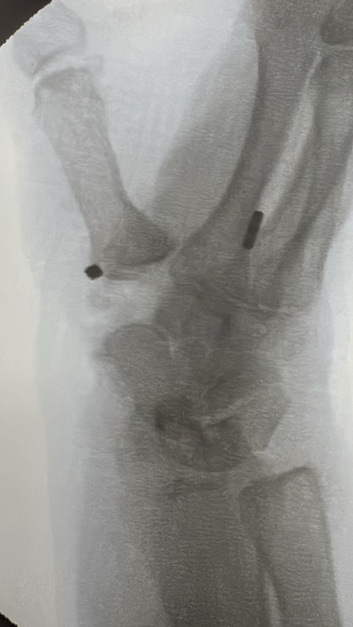
The anatomy at the base of the thumb is different from any other part of the body. The trapezium bone, which articulates with the 1st metacarpal, has two other surface areas with the scaphoid and the trapezoid. If there is arthritis between the scaphoid and the trapezium, it is best to do a procedure called a Trapeziectomy. In this procedure, the trapezium is removed thereby eliminating the bone-on-bone contact between the 1st metacarpal and the scaphoid, which had created pain and discomfort. The trapezium has no more purpose and removing it solves two problems of STT (scapho-trapezium-trapezoid) arthritis and CMC arthritis. A suspensionplasty is done with an implant, called a tightrope. The tightrope suspends the first to the second metacarpal, preventing the first metacarpal from falling into the empty space where the trapezium was. This is an excellent procedure, restoring the position of the thumb and assists the body to regain muscle strength once the metacarpal is placed in its normal anatomical position and it can move freely without pain.
In summary, in isolated CMC arthritis, especially in the younger patients, a CMC joint replacement is an excellent treatment solution. However, if the STT joint is involved, a trapeziectomy with a suspensionplasty provides excellent outcomes with regaining of strength of the thumb and the ability to return to normal activities.
Please read the above text for more in-depth information to help answer these questions. It's important to consult with our qualified healthcare professionals to ensure the appropriate diagnosis and treatment plan for your specific situation.